
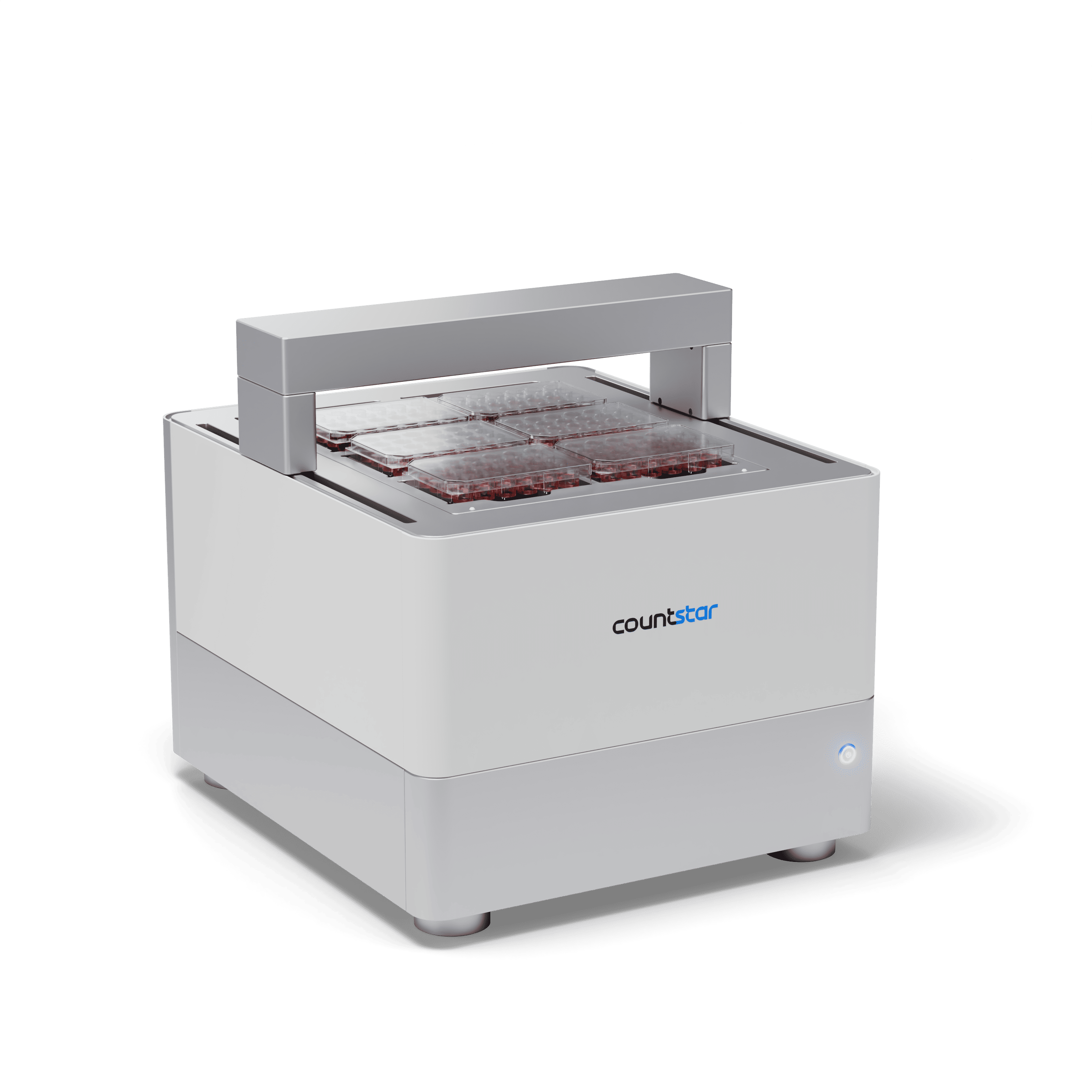
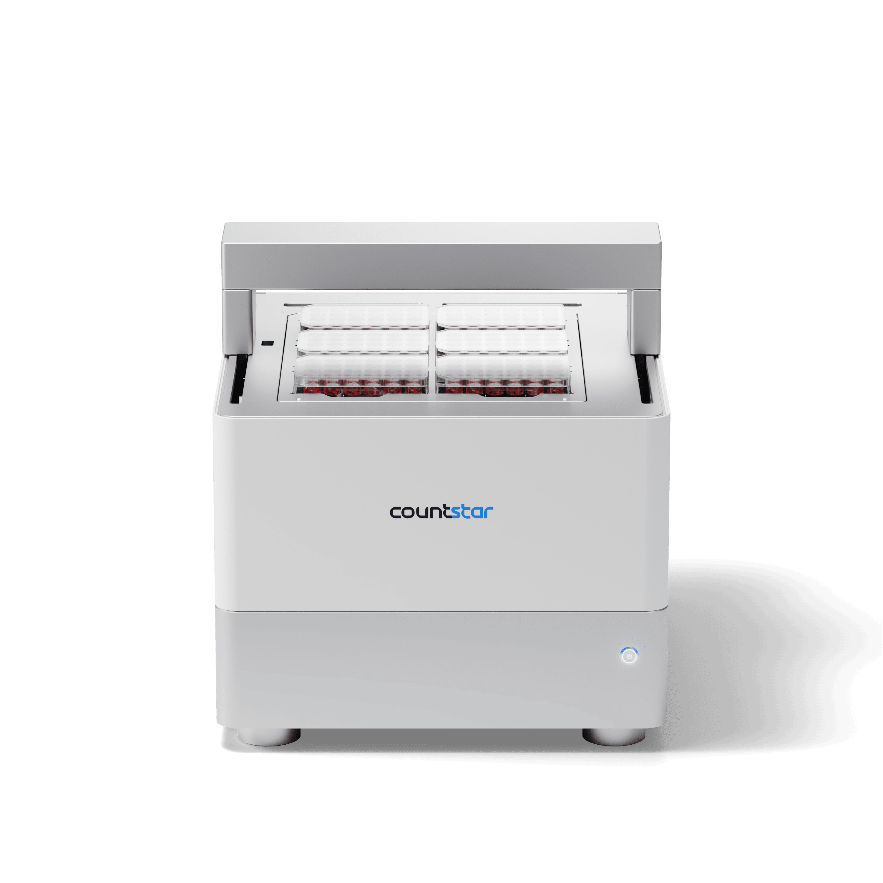

Spica M6
Turn Biological Curiosity into Kinetic Clarity.
Live Cell Analysis
High Throughput
In-Incubator System
6-plate configuration
Fluorescence
Mammalian Cells
Advanced Cell Models
Cell Health & Quality Control
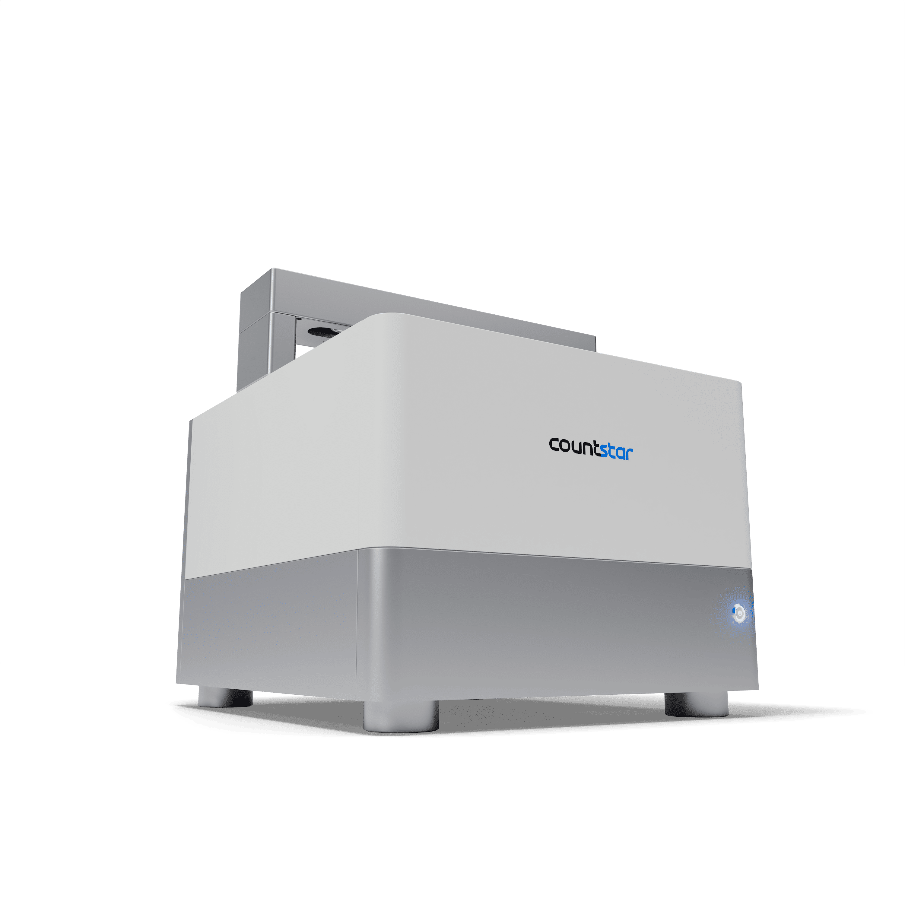
Meet the Spica M6. A 6-plate, AI-driven platform that tracks your live-cell assays from Day 0 to definitive discovery—right inside your incubator.
Elevate your live-cell analysis workflow
The Spica M6 eliminates the bottlenecks of kinetic analysis. We combined motorized 6-plate walk-away automation with high-speed, 4-channel optics to capture the complete dynamic narrative of your cells—without ever disturbing their environment.
Native Incubator Integration
Operates completely inside standard incubators to preserve absolute physiological integrity.
True 6-Plate Walk-Away Scale
Maximize throughput with an automated 6-plate deck, allowing for independent imaging schedules and seamless adaptation to flasks and dishes.
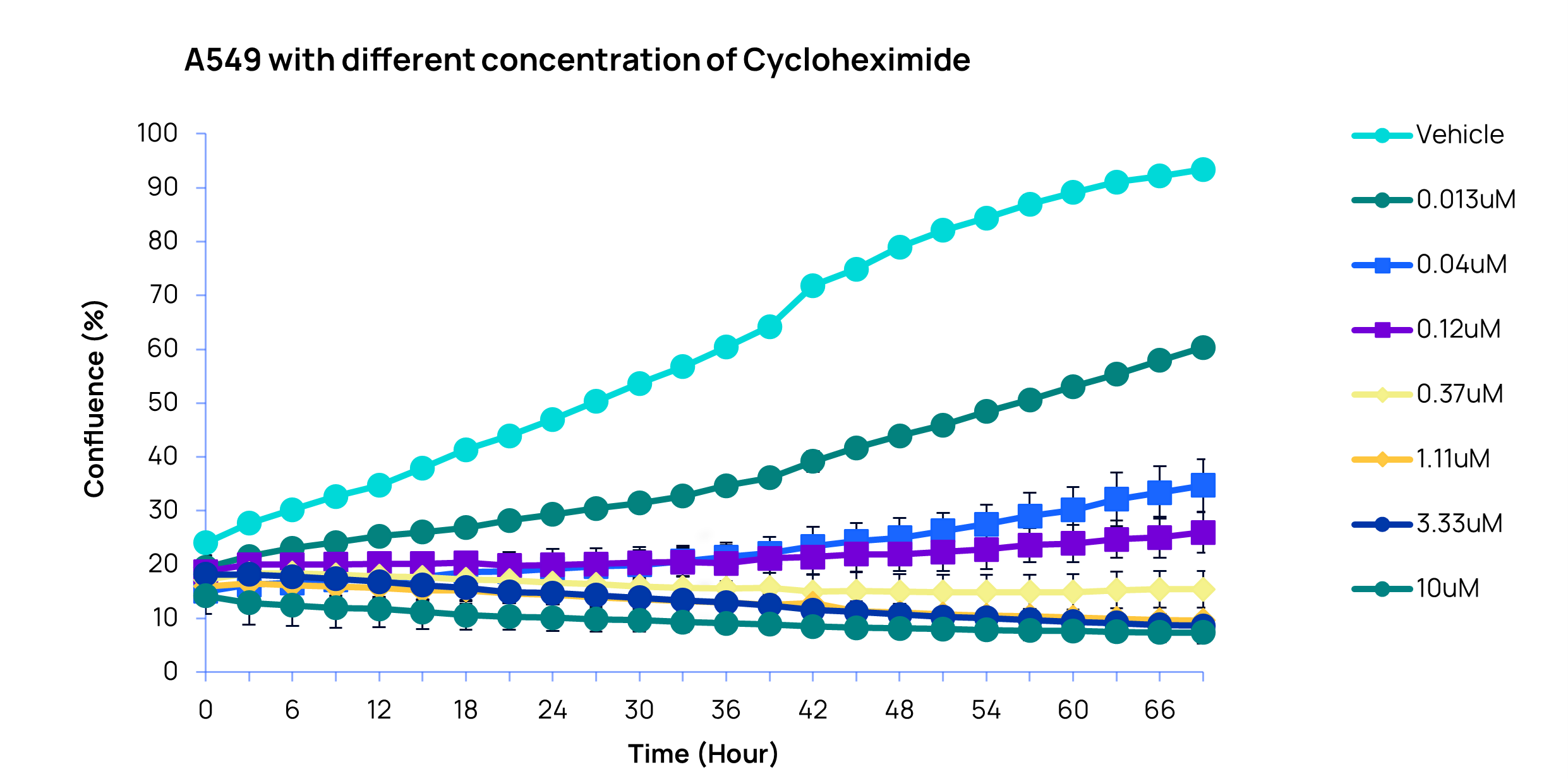
Real-Time Kinetic Analysis
Generate presentation-ready curves and actionable metrics long before the run is complete.
Adaptive Dual-Engine Intelligence
Toggle seamlessly between rapid routine assays and advanced, bias-free AI modeling.
4-Channel Multiparametric Optics
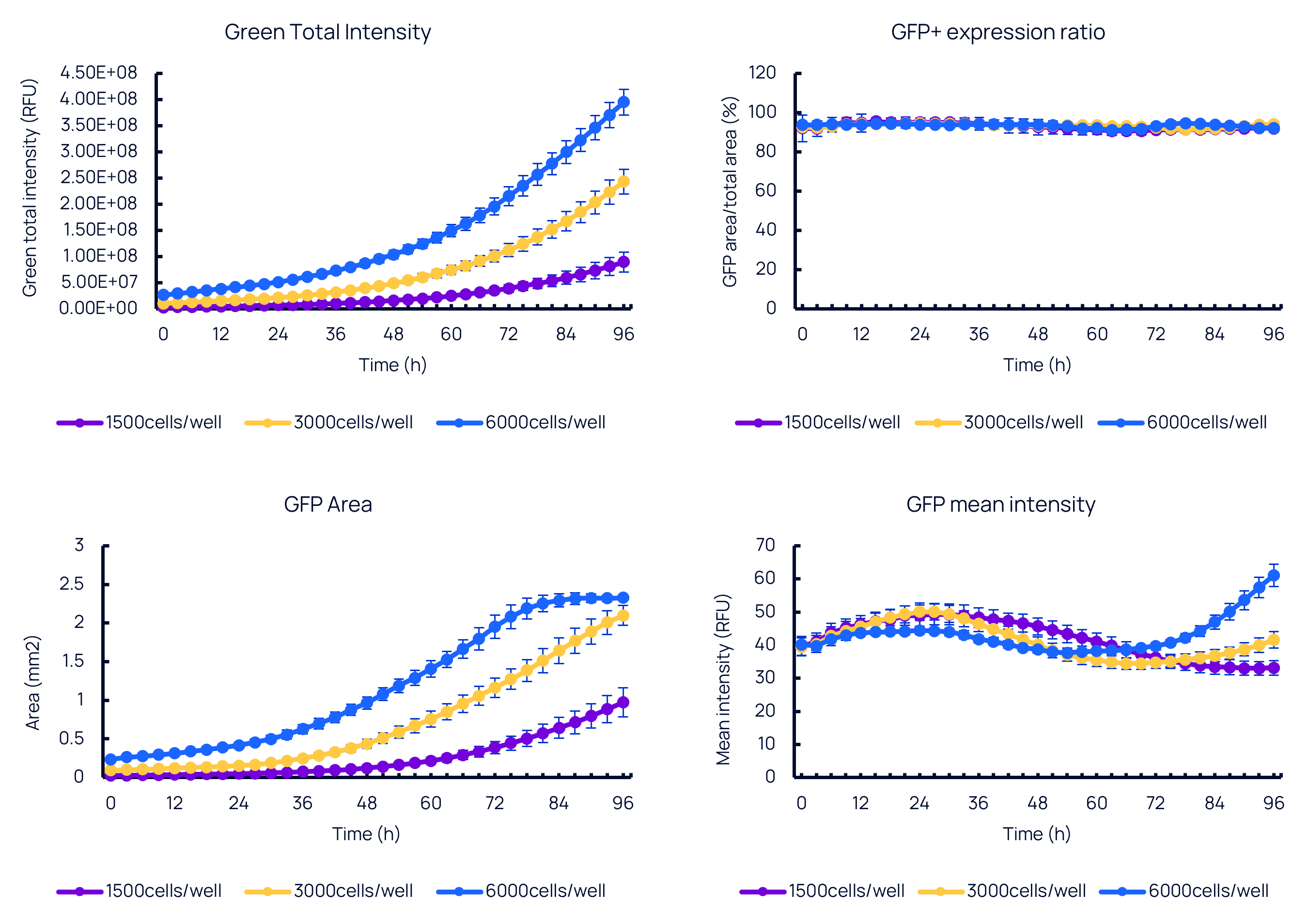
Track proliferation, apoptosis, and spatial morphology simultaneously within a single well.
Networked Multi-User Workspace
Enable multiple users to concurrently monitor assays, schedule scans, and export results from anywhere on your local network.
Product Details
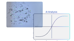
Transparent AI. No Black Boxes.
Traditional imaging software locks you into rigid algorithms. The Spica M6 utilizes an adaptive, two-tiered software ecosystem that puts you in control.
The Standard Engine:
Rapid, one-click quantification for routine workflows like confluence and cell health assays
The Stellar AI Layer:
Advanced, no-code customization. Easily train bespoke models and define tailored metrics across your entire 6-microplate matrix.
Learn more about Stellar AI
Engineered for Precision
Beyond 6-plate walk-away automation, the Spica M6 optical drive is built to capture every biological nuance without compromise.
Minimal Cell Disruption:
Optics move beneath the deck while plates remain stationary, eliminating fluid shear stress to protect delicate cultures.
High-Multiplex Fluorescence:
Cover a broad spectral range to support diverse reagents. Access up to 5 distinct colors, multiplexing a maximum of 4 simultaneously.
Automated Turret (4x, 10x, 20x):
Switch magnifications for individual assay—from full-well 4x overviews to high-resolution 20x subcellular tracking.
User-Defined Z-Stacking:
Set precise focal planes to capture custom z-stacks, keeping complex 3D organoids in perfect focus.
Applications
Uncover the Full Kinetic Story in Your Research
Oncology & Immuno-Oncology
Visualize complex tumor microenvironments and immune responses over time.
Key Assays:
Immune Cell Killing
Phagocytosis & Efferocytosis
Apoptosis & Cytotoxicity.
Drug Discovery & Screening
Track multiplexed dose-response assays to confidently identify lead compounds earlier.
Key Assays:
Dose-Response Kinetics
Cell Cycle Dynamics
Antibody Internalization
3D Biology & Organoids
Track the growth, viability, and differentiation of complex organoids over weeks.
Key Assays:
Organoid Growth & Viability
3D Morphological Profiling
Tumor Spheroid Invasion
Cell Line Development & Optimization
Streamline generation and maintenance with continuous proliferation tracking.
Key Assays:
Clonal Outgrowth & Confluence
Reporter Gene Expression
Transfection Efficiency
Stem Cell Research
Quantify lineage commitments and expansion rates as your cultures mature.
Key Assays:
Pluripotency Marker Expression
Embryoid Body (EB) Formation
Stem Cell Expansion
Cell & Gene Therapy
Continuously monitor target engagement to validate the persistence of engineered therapies.
Key Assays:
CAR-T Cell Expansion
Immune Cell Clustering & Target Engagement
Transduction Efficiency
Tech Specs
Specifications & related products
Category
Specification
Details
Camera
High-sensitivity CMOS camera
2200 * 3200
Fluorescence Channel
Blue / Green / Orange / Far Red
Ex=369-414,Em=415-455|Ex=456-500,Em=501-537|Ex=537-571,Em=572-615|Ex=616-658,Em=659-736
Blue / Green / Red
Ex: 359-400, Em: 408-440Ex: 450-489, Em: 500-546Ex: 555-605, Em: 615-705
Optical Magnification
4x
Resolution: 1.13μm/pixelField of View: 3.6 × 2.48mm
10x
Resolution: 0.45μm/pixelField of View: 1.44 × 0.99mm
20x
Resolution: 0.23μm/pixelField of View: 0.72 × 0.50mm
Spica M6 Imaging Unit
Dimensions (W × L × H)
428 × 498 × 450mm
Operating Conditions
10-40°C, RH up to 95%
Focus Mode
Image-based focus
Imaging Mode
Bright-field & fluorescence microscopy, Z-axis layer scanning
6-96 whole well, Z-stack range > 1mm
Supported Consumables
6-384 well plates
35mm/ 60mm / 100mm dishes, T25/ T75/ T175 flasks
Analysis Algorithms
AI analysis, conventional image processing
Export Format
Image
JPEG/ TIFF/ BMP/ PNG
Video
AVI/ MP4
Data
XLSX/ CSV
Ready to
Upgrade your Live Cell Analysis?